

Diagnosis of vitiligo
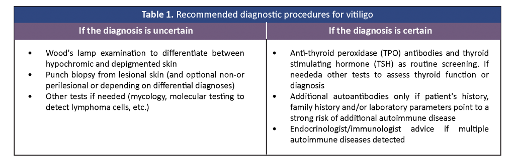
Vitiligo is characterized by the progressive loss of melanocytes, leaving white patches on the skin. It is usually diagnosed by clinical examination alone supported by Wood’s lamp examination. The differential diagnoses of vitiligo include pityriasis alba, hypopigmented mycosis fungoides, tinea versicolor, idiopathic guttate hypomelanosis, and other hypo-or depigmented disorders. Additionally, the diagnosis of vitiligo could be wrongly made in patients with fair skin who developed due to the intervening normal skin within the melasma macules giving a vitiligo-like appearance. In cases of uncertain diagnosis, a skin biopsy, mycologic examination and appropriate blood tests may be needed to exclude a fungal infection, cutaneous lymphoma and other disorders. Routine screening of anti-thyroid antibodies and thyroid function is recommended. If the patient’s history or routine laboratory parameters suggest additional autoimmune disorders, further investigation and management are strongly suggested (Table 1).
Management of vitiligo
The experts developed a management algorithm to guide the assessment, decision-making and treatment of vitiligo (Figures 1 and 2).
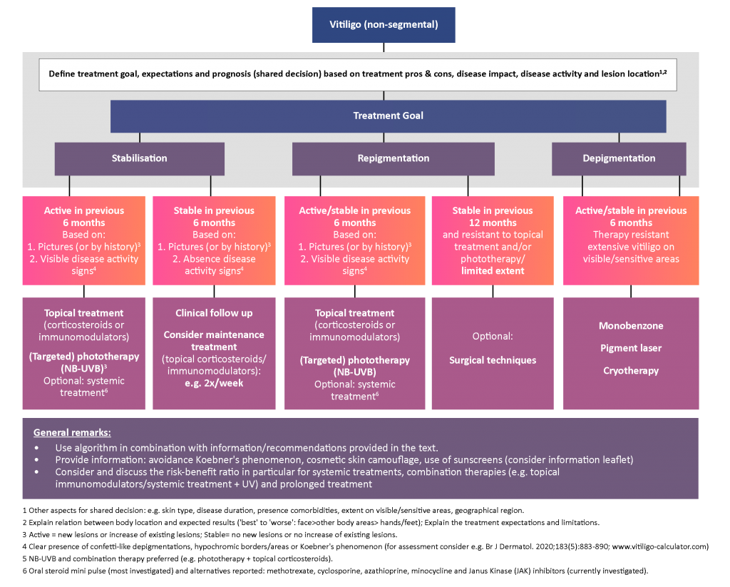
Figure 1. Recommendations for the management of vitiligo (non-segmental)
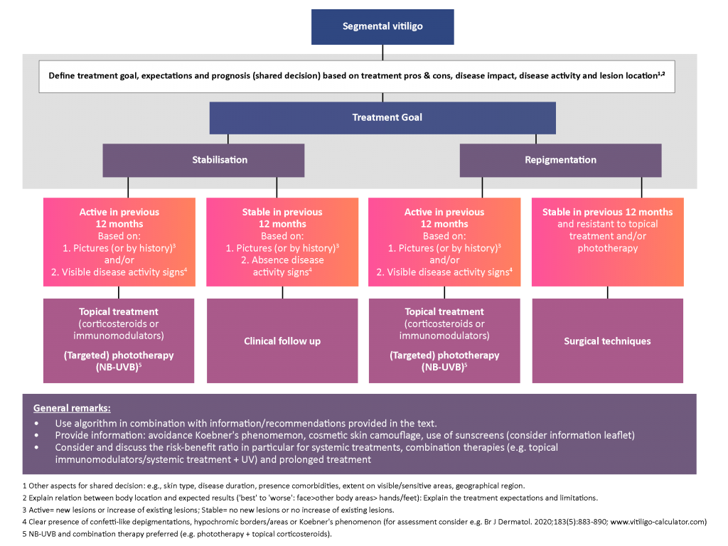
Figure 2. Recommendations for the management of segmental vitiligo
Reference: van Geel N, Speeckaert R, Taïeb A, et al. Worldwide expert recommendations for the diagnosis and management of vitiligo: Position statement from the International Vitiligo Task Force Part 1: Towards a new management algorithm. J Eur Acad Dermatol Venereol. 2023;37(11):2173–2184.