

Melasma is a common, chronic, and relapsing hyperpigmentation disorder caused by overactive melanocytes that produce and deposit excessive melanin in the epidermis and dermis. It is especially prevalent in females, particularly those of reproductive age, and commonly occurs in areas frequently exposed to sunlight, such as the face.1
Melasma typically appears as irregular, light brown to gray-brown patches and macules on sun-exposed areas. These lesions are generally symmetrical and commonly affect the forehead, nose, cheeks, upper lip, and chin.¹
In facial melasma, three typical patterns of distribution are observed: The centrofacial pattern, which involves the forehead, cheeks, nose, upper lip, and chin¹ (Figure 1)²; the malar pattern, mainly affecting the lateral cheeks¹ (Figure 2)²; and the mandibular pattern, which appears along the lower jawline¹ (Figure 3).²
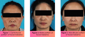
Some patients, mainly females, develop extrafacial melasma, a less common form compared to the facial type, and it is typically more challenging to manage.¹
Differential diagnosis of melasma
The differential diagnosis of melasma encompasses a wide range of disorders characterized by facial hyperpigmentation and includes the following.
Hori’s nevus
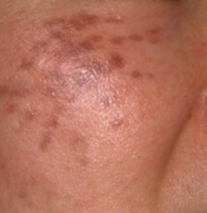
Distinguishing characteristics: Numerous speckled blue-brown or slate-grey macules appearing simultaneously on both malar regions.1
Riehl’s melanosis
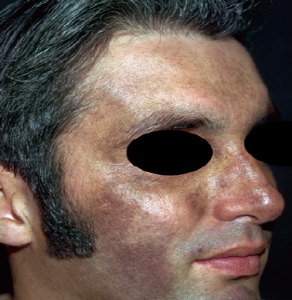
Distinguishing characteristics: Brown-grey reticulated-to-diffuse hyperpigmented macules or patches on the face, neck, and upper chest4
Erythema dyschromicum perstans
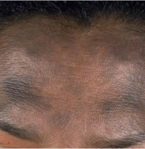
Distinguishing characteristics: Hyperpigmented macules that are well-circumscribed round to oval or irregular patches on the face, neck and trunk, and grey in color1,5
Lichen planus pigmentosus
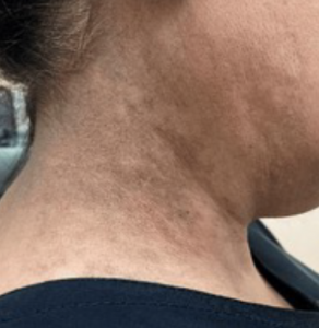
Distinguishing characteristics: Oval or irregular, brown to grey macules and patches most often seen in sun-exposed areas of the skin, such as the face and flexural areas1
Fixed drug eruptions
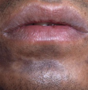
Distinguishing characteristics: Solitary, round to oval, dusky red to brown/black macules that re-occur in the same regions when re-exposed to the causative drug.1
Discoid lupus erythematosus
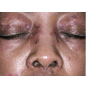
Distinguishing characteristics: Presents with violaceous and post inflammatory hyperpigmentation, interspersed with hypopigmented, scar-like areas.1
Phototoxic dermatitis
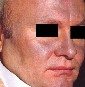
Distinguishing characteristics: Exaggerated sunburn confined to sun-exposed areas, often followed by post-inflammatory hyperpigmentation.1
E.g., Slate grey pigmentation in a photodistributed pattern is produced on the face by amiodarone.1
Phytophotodermatitis
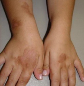
Distinguishing characteristics: Hyperpigmented macules or patches with irregular shapes that match the areas of contact with sensitizing substances, such as lemons.1
Lentigines
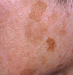
Distinguishing characteristics: Multiple tan to dark brown macules, often with irregular borders1
Melasma is an acquired hyperpigmentation disorder marked by light to dark brown irregular macules or patches on sun-exposed skin, especially the face. Despite its frequency and treatment demand, diagnosis can be challenging due to similar-looking conditions. Hence, dermatologists must consider common differentials when evaluating melasma.8
References:
- Melasma: Epidemiology, pathogenesis, clinical presentation, and diagnosis. Available at: https://pearlgrimesmd.com/wp-content/uploads/2023/09/Melasma_-Epidemiology-pathogenesis-clinical-presentation-and-diagnosis-UpToDate.pdf. Accessed on July 10, 2025.
- Zheng H, Pei Q, Yao M. Understanding melasma: From pathogenesis to innovative treatments. Dermatologic Therapy. 2024;1:1–9.
- Horis Nevus treatment. Available at: https://apaxmedical.com/horis-nevus-treatment/. Accessed on July 10, 2025.
- Riehl melanosis. Treasure Island (FL): StatPearls Publishing; 2025 Jan-. Available at: https://www.ncbi.nlm.nih.gov/books/NBK557437/. Accessed on July 10, 2025
- Erythema dyschromicum perstans. Available at: https://dermnetnz.org/topics/erythema-dyschromicum-perstans. Accessed on July 10, 2025.
- Almarek RA, AlQadri NG, Alotaibi M. Coexistence of lichen planus pigmentosus and classic lichen planopilaris: A case report and literature review. Cureus. 2023;15(10):e46952.
- Available at: https://www.aocd.org/page/Phytophotodermatitis. Accessed on July 10, 2025.
- Honigman A, Rodrigues M. Differential diagnosis of melasma and hyperpigmentation. Dermatological Reviews. 2023;4:30–37.
Expert opinion – Dr. Vinayak Venkatesh
When evaluating a patient with suspected melasma, it is important to keep in mind that several other conditions can present with a similar appearance. Apart from the conditions explained above, other conditions that mimic melasma include post-inflammatory hypermelanosis, actinic lichen planus, minocycline-induced pigmentation, poikiloderma of Civatte, and exogenous ochronosis from long-term hydroquinone use.
Diagnosis can be confirmed through thorough clinical examination, use of Wood’s lamp, dermoscopy, and, when required, skin biopsy.