

Pityriasis rosea, also known as pityriasis circinata, roseola annulata, and herpes tonsurans maculosus, is an acute self-limiting papulosquamous disorder. PR is characterized clinically by a herald patch, or mother patch, followed by scaly oval plaques on the trunk and proximal extremities along the Langer lines of cleavage, giving a characteristic “Christmas tree” appearance. Collarette scaling is common (Figure 1). Pruritus is severe in 25% of cases. The herald patch is seen in 50% to 90% of patients and is located on the trunk, followed by the neck or proximal extremity. Over 1 to 2 weeks after the onset of the herald patch, a generalized skin eruption occurs, in which numerous lesions develop in crops. The eruption is usually preceded by a prodrome of a sore throat, gastrointestinal disturbance, fever, and arthralgia. The resulting lesions are symmetric and most commonly involve the thorax, back, abdomen, and adjoining areas of the neck and extremities. The condition commonly resolves spontaneously within weeks to a few months. Symptomatically, mild-to-moderate itching might accompany the rash, but the overall impact on health is minimal for most individuals. Recurrence is rare (<5%), with most cases presenting as a singular occurrence in a person’s lifetime.1
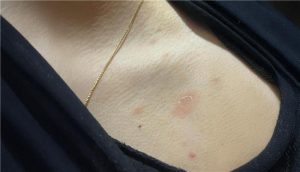
Differential diagnosis of pityriasis rosea
Seborrheic dermatitis
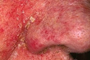
Distinguishing characteristics: Orange-red or gray-white skin with greasy or white dry scaling macules, papules, or patches; diffuse scalp involvement with marked scaling; worsens in winter because of dry conditions; pruritus increases with perspiration2
Secondary syphilis
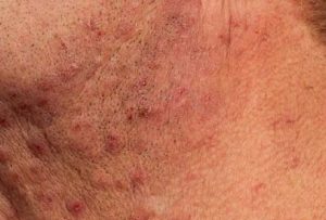
Distinguishing characteristics: 0.5- to 1-cm, pink to brownish-red, round to oval macules and papules on the trunk, palms, and soles; patchy, “moth-eaten” alopecia on the scalp and beard area; mucous membrane involvement with round or oval patches covered by hyperkeratotic white to gray membrane2
Tinea corporis
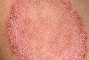
Distinguishing characteristics: Scaling, sharply marginated plaques of various sizes with or without pustules or vesicles along the margins; lesions present with peripheral enlargement and central clearing, producing an annular configuration with concentric rings or arcuate lesions2
Viral exanthems
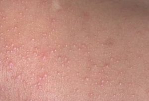
Distinguishing characteristics: Diffuse maculopapular erythema; mucosal involvement with microulcerative lesions, palatal petechiae, or conjunctivitis; systemic findings of lymphadenopathy, hepatomegaly, and splenomegaly2
Pityriasis rosea is a common self-limiting rash that usually starts with a herald patch on the trunk and progresses along the Langer lines to a generalized rash over the trunk and limbs. The diagnosis is based on clinical and physical examination findings. The herald patch is an erythematous lesion with an elevated border and depressed center.2
Reference:
- Litchman G, Nair PA, Syed HA, et al. Pityriasis Rosea. [Updated 2024 Mar 1]. In: StatPearls [Internet]. Treasure Island (FL): StatPearls Publishing; 2025 Jan-. Available from: https://www.ncbi.nlm.nih.gov/books/NBK448091/.
- Villalon-Gomez JM. Pityriasis rosea: Diagnosis and treatment. Am Fam Physician. 2018;97(1):38–44.
Expert opinion – Dr. Kavya K
Pityriasis rosea is among the most common papulosquamous disorders seen in dermatology. Patients typically present with mild, scaly lesions predominantly over the trunk, arms, and back. The condition often begins with a single, larger scaly plaque known as the herald patch or mother patch, usually located on the trunk or proximal upper extremities. Within 1–2 weeks, multiple smaller lesions appear, classically distributed in a “Christmas-tree” pattern on the trunk and upper back. Most patients are asymptomatic or experience mild pruritus, and some may report viral prodromal symptoms preceding the eruption. Although pityriasis rosea is generally self-limiting, resolution may take several weeks to months, with recurrence occurring in about 5% of individuals. Diagnosis is usually clinical, though it can mimic seborrheic dermatitis, tinea corporis, or secondary syphilis. Treatment is mainly supportive, involving emollients and mild topical corticosteroids, with improvement typically seen within 1–2 weeks in symptomatic cases.