

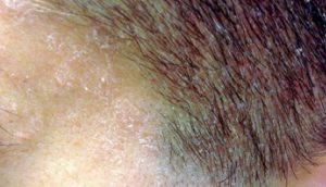
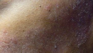
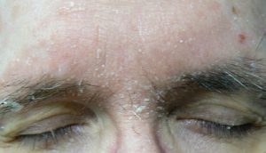
Seborrheic dermatitis is a chronic inflammatory dermatologic condition that usually appears on areas of the body with a large density of sebaceous glands, such as the scalp, face, chest, back, axilla, and groin. Seborrheic dermatitis is a clinical diagnosis based on lesion location and appearance. In infants, it may present as thick white or yellow greasy scales on the scalp; it is usually benign and resolves spontaneously. In adolescents and adults, seborrheic dermatitis typically presents as flaky, greasy, erythematous patches on the scalp (Figure 1), nasolabial folds (Figure 2), ears, eyebrows (Figures 3), anterior chest, or upper back. Although the pathophysiology of seborrheic dermatitis is not completely understood, the mechanisms of effective therapies coupled with results of recent biomolecular studies provide clues about the causes. The redness, itching, and scaling associated with seborrheic dermatitis are caused by changes in skin cell functioning. Malassezia yeast seems to cause a nonspecific immune response that begins the cascade of skin changes that occur in seborrheic dermatitis.
Differential diagnosis of seborrheic dermatitis
Lichen simplex chronicus
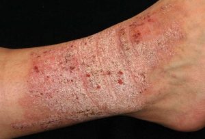
Distinguishing characteristics: Eczematous eruption caused by habitual scratching of a single localized area; more common in adults, but possible in children
Secondary syphilis
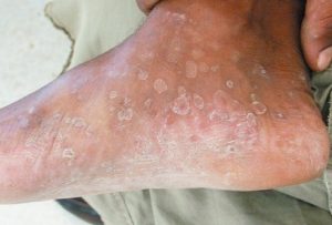
Distinguishing characteristics: Copper-colored scaly plaques on palms and soles accompanied by an influenza-like syndrome and generalized adenopathy
Systemic lupus erythematosus
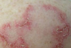
Distinguishing characteristics: Discoid: disk-like lesions on face and scalp; subacute: photo-distribution papulosquamous, annular lesions over trunk; acute: photo-distribution red plaques forming facial butterfly rash; infants: appears in first month of life; papulosquamous, annular lesions
Psoriasis
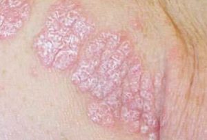
Distinguishing characteristics: Distinctive red, scaling papules that coalesce to form round-to-oval plaques
The differential diagnosis is lengthy, but the correct diagnosis can usually be made clinically by the characteristic distribution of lesions and varying course of the disease. If the diagnosis is uncertain, a biopsy demonstrating parakeratosis in the epidermis, plugged follicular ostia, and spongiosis can confirm the presence of seborrheic dermatitis.
Reference:
- Clark GW, Pope SM, Jaboori KA. Diagnosis and treatment of seborrheic dermatitis. Am Fam Physician. 2015;91(3):185–190.
Expert opinion – Dr. Shruthi Reddy
The article gives a clear and concise explanation of seborrheic dermatitis, describing its appearance, common locations, and how it differs from similar skin conditions. The information is easy to understand and scientifically accurate. However, the article could be even more useful if it included brief treatment or management tips. Overall, it is an informative and well-organized piece. Often only SD and scalp psoriasis is confused but the type of scales and distribution helps to make the diagnosis.