

Tinea versicolor, also known as pityriasis versicolor, is a common, benign, superficial fungal skin infection. It is caused by saprophytic, lipid-dependent yeast from the genus Malassezia, formerly known as Pityrosporum, which is part of the normal skin flora. Unlike other forms of tinea, tinea versicolor is not a dermatophyte infection. Clinical features include hyperpigmented or hypopigmented macules with fine overlying scales. The most affected areas are the trunk, neck, and proximal extremities. Diagnosis of pityriasis versicolor is usually clinical. When uncertain, a Wood’s lamp examination and microscopic analysis of scales soaked in potassium hydroxide can be helpful. Medical treatment is effective for tinea versicolor; however, the condition often recurs, so that ongoing preventive measures may be necessary over an extended period.
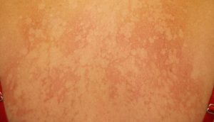
The characteristic feature of tinea versicolor is multiple, well-demarcated, oval, finely scaling patches or plaques. Skin lesions may be hypopigmented, hyperpigmented, or erythematous and occasionally become confluent and widespread (Figure 1). Hyperpigmented tinea versicolor is typically a light brown in those with a fair complexion. In patients with darkly pigmented skin, hyperpigmented tinea versicolor can manifest as macules and patches ranging from dark brown to grayish black. Affected areas can vary in hue within the same person and differ among people of the same skin tone. The underlying scale may not be readily apparent on the lesions, but stretching or scraping the affected skin readily provokes it, a sign known as the “evoked scale sign.”
Differential diagnosis of tinea versicolor
Pityriasis rosea
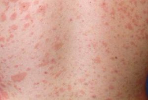
Distinguishing characteristics: Inflammatory macules surrounded by a collarette of scales on the trunk in a “Christmas tree-like” pattern. Often, a bigger herald patch manifests before the general eruption
Erythrasma
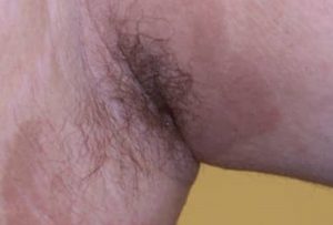
Distinguishing characteristics: Erythematous or hyperpigmented regions in the axillae or groin
Vitiligo
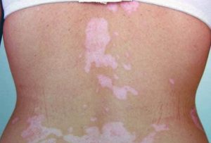
Distinguishing characteristics: Completely depigmented macules and patches
Guttate psoriasis
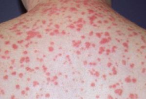
Distinguishing characteristics: Small, erythematous, scaling plaques affecting primarily the trunk and upper arms
Recognizing the characteristic color variation and evoked scale of tinea versicolor is essential, as several conditions—such as pityriasis rosea, erythrasma, vitiligo, and guttate psoriasis—can closely resemble its macular presentation. Careful clinical evaluation helps differentiate these look-alike disorders and ensures accurate diagnosis and appropriate management.
Reference:
- Karray M, McKinney WP. Tinea Versicolor. [Updated 2024 Feb 12]. In: StatPearls [Internet]. Treasure Island (FL): StatPearls Publishing; 2025 Jan-. Available from: https://www.ncbi.nlm.nih.gov/books/NBK482500/.
Expert opinion – Dr. Abhishek Shetty
This article provides a crisp yet thorough overview of tinea versicolor as commonly encountered in daily practice, with a particularly useful emphasis on pigment variation across different skin types. The reminder about the evoked scale sign is valuable, as it remains one of the most reliable clinical clues. I find the differentiation from pityriasis rosea and erythrasma especially relevant, since these conditions often mimic early presentations. The clarification regarding complete depigmentation in vitiligo reinforces an important distinction that patients frequently misunderstand. The mention of guttate psoriasis adds practical value, especially in younger patients presenting with truncal lesions. The discussion on frequent recurrence aligns well with real-world experience and highlights the importance of long-term maintenance therapy. The write-up also reiterates that diagnosis is largely clinical, with Wood’s lamp examination and KOH testing reserved for atypical cases. Its attention to color variability in darker phototypes accurately reflects what we commonly observe in diverse populations.