

Melasma is an acquired hyperpigmentation disorder characterized by light-to-dark brown-colored irregular macules or patches on sun-exposed areas of the skin, typically the face. It is the most common cause of facial pigmentation and is a cutaneous disorder affecting all races with particular prominence in darker-skinned individuals. Melasma presents clinically as brown macules or patches and is generally a clinical diagnosis. The most common clinical pattern (50%–80% of cases) is the centrofacial pattern, where lesions are predominantly in the center of the face – affecting the forehead, cheeks, nose, upper lip, or chin (Figure 1). Epidermal melasma is the most common form and is characterized by pigment deposition throughout the layers of the epidermis, particularly in the basal and suprabasal layers, and occasionally extending throughout the entire epidermis. Melanocytes in the epidermis are generally enlarged, have prominent dendrites, and increased melanosomes. Dermal melasma exhibits pigment deposition throughout the epidermis and the upper and middle layers of the dermis, occasionally extending into the deep dermis.
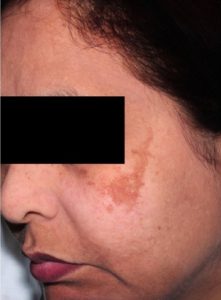
Differential diagnosis of melasma
Café-au-lait macules
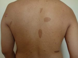
Distinguishing characteristics: They are asymptomatic and present as light-to-dark brown well-circumscribed, pigmented macules or patches usually greater than 20 mm in size and often located on the trunk. They are epidermal in origin, representing an increase in melanin in melanocytes and basal keratinocytes.
Post-inflammatory hyperpigmentation
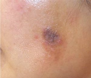
Distinguishing characteristics: Hyperpigmentation within the dermis has a blue–gray appearance and may either be permanent or resolve over a prolonged period of time if left untreated.
Exogenous ochronosis
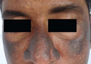
Distinguishing characteristics: It manifests as hyperpigmentation in photo-exposed regions, often affecting the zygomatic regions in a symmetrical pattern. The lesions are typically blue–black macules, usually accompanied by pinpoint and caviar-like papules, and are frequently confused with melasma, PIH, and Riehl’s melanosis.
Acanthosis nigricans
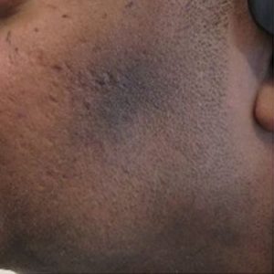
Distinguishing characteristics: Velvety hyperpigmented macules and patches that progress to symmetrical palpable plaques
Melasma and other causes of facial hyperpigmentation affect millions of people worldwide. Often, these conditions can be difficult to differentiate and diagnose, which may delay correct and timely management. This may lead to disease progression, incorrect treatment, and worsening of the condition. Furthermore, these conditions often have a negative impact on both physical and emotional well-being. Therefore, clinicians must have comprehensive knowledge and a systematic approach to melasma and its common differential diagnoses.
Reference:
- Hongiman A, Rodrigues M. Differential diagnosis of melasma and hyperpigmentation. Dermatological Reviews. 2023;4:30–37.
Expert opinion – Dr. Manjunath J V
Melasma is the most common facial pigmentation disorder encountered in clinical practice, and diagnosis is typically made clinically. This article provides a clear overview to help differentiate melasma from other causes of facial hyperpigmentation, making it highly valuable for everyday practice. I would also like to emphasize that the various subtypes of melasma can themselves present diagnostic challenges. Distinguishing between epidermal and dermal melasma is important for guiding appropriate treatment, and a Wood’s lamp examination can aid in assessing pigment depth. Overall, this is a well-written and practically useful article for clinicians.