

Seborrheic dermatitis (SD) is a common papulosquamous skin disease occurring predominantly in infancy and middle age, exhibiting distinct variations across these age groups. Characterized by folliculocentric salmon-colored papules and plaques with a fine white scale and a yellowish crust (often described as a greasy scale-crust), SD manifests diversely across different body areas and may present in multiple locations (Figure 1). Because it often resembles other dermatoses, SD necessitates accurate differentiation. Notably, flexural surfaces typically display lesser scaling, with poorly defined margins. The dermatopathology of SD is nonspecific, but the surface and infundibular epidermis usually show a superficial perivascular infiltrate of lymphocytes, acanthosis, focal spongiosis, and focal parakeratosis.1
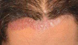
Differential diagnosis of Seborrheic dermatitis
Atopic dermatitis
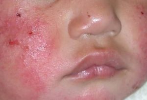
Distinguishing characteristics: Flexural lichenification in adults; facial and extensor involvement in infants and children2
Impetigo
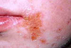
Distinguishing characteristics: Superficial skin infection caused by streptococci and/or staphylococci; begins as vesicles with thin, fragile roof2
Pityriasis rosea
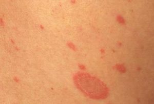
Distinguishing characteristics: Begins with herald patch; “Christmas-tree distribution” of salmon pink papules over trunk and proximal extremities2
Tinea capitis, corporis
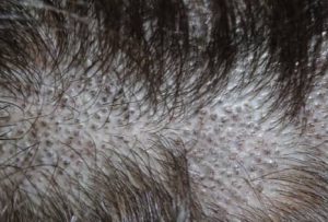
Distinguishing characteristics: Dermatophyte infection of the scalp or body; leading edge (active border) scaly, red, and slightly elevated with central clearing; vesicles appear at the active border when inflammation is intense; classic “ringworm pattern”2
Seborrheic dermatitis can closely resemble several skin disorders, but recognizing its characteristic greasy scale, distribution, and clinical patterns ensures accurate diagnosis and effective management.
Reference:
- Tucker D, Masood S. Seborrheic Dermatitis. [Updated 2024 Mar 1]. In: StatPearls [Internet]. Treasure Island (FL): StatPearls Publishing; 2025 Jan-. Available from: https://www.ncbi.nlm.nih.gov/books/NBK551707/
- Clark GW, Pope SM, Jaboori KA. Diagnosis and treatment of seborrheic dermatitis. Am Fam Physician. 2015;91(3):185–190.
Expert opinion – Dr. Ravi H B
Seborrheic dermatitis remains one of the most frequently encountered papulosquamous disorders in clinical practice; however, its clinical overlap with several inflammatory and infectious dermatoses continues to challenge diagnostic accuracy, even among experienced clinicians. Classically, seborrheic dermatitis presents with folliculocentric, salmon-colored papules and plaques accompanied by fine white scaling and a greasy yellow crust, predominantly affecting sebaceous-rich areas such as the scalp, face, chest, and flexures, where margins may appear ill-defined, particularly in intertriginous regions. Differentiation from atopic dermatitis is aided by the presence of flexural lichenification in adults and facial or extensor involvement in children, typically without the greasy scale characteristic of seborrheic dermatitis. Impetigo must be excluded when erosions and prominent yellow crusting are present, especially if fragile vesicles or signs of bacterial colonization are noted. Pityriasis rosea is distinguished by an initial herald patch followed by a characteristic Christmas-tree distribution over the trunk and proximal extremities, while tinea infections are suggested by active borders, central clearing, and occasional vesiculation, with potassium hydroxide testing providing confirmation when uncertainty persists. In infants, seborrheic dermatitis commonly manifests as cradle cap with thick greasy scales, whereas adults often experience chronic, relapsing involvement of the scalp and face. Histopathology is nonspecific, typically showing acanthosis, spongiosis, parakeratosis, and a superficial lymphocytic infiltrate. Ultimately, diagnosis relies on anatomical distribution, the quality of scaling, chronicity, and the absence of primary vesiculation, with early recognition of clinical mimickers being essential to prevent mismanagement and ensure appropriate, targeted therapy, particularly in recurrent or treatment-resistant cases.