

Lichen planus is a chronic, inflammatory, autoimmune disease that affects the skin, oral mucosa, genital mucosa, scalp, and nails. Lichen planus lesions are described using the six P’s (planar [flat-topped], purple, polygonal, pruritic, papules, plaques). Onset is usually acute, affecting the flexor surfaces of the wrists, forearms, and legs. The lesions may appear in a linear configuration, following the lines of trauma. It is common to see post-inflammatory hyperpigmentation as the cutaneous lesions clear, especially in persons with darker skin. Cutaneous lichen planus may present in different forms. Linear lichen planus manifests as closely aggregated linear lesions on the limbs that may develop the Koebner phenomenon. Annular lichen planus (Figure 1) accounts for approximately 10% of lichen planus cases. It commonly appears as arcuate groupings of individual papules that develop rings or a peripheral extension of clustered papules with central clearing. In addition to the usual sites of distribution, this form of lichen planus may occur on male genitalia and buccal mucosa.
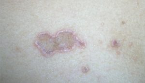
Differential diagnosis of cutaneous lichen planus
Eczema
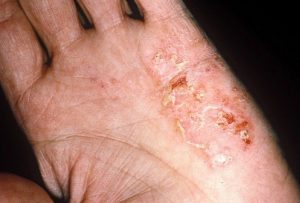
Distinguishing characteristics: Excoriations and lichenification of skin, often on flexor surfaces
Lichen simplex Chronicus
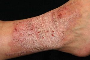
Distinguishing characteristics: One or more plaques with lichenification in an area that is easily scratched
Pityriasis rosea
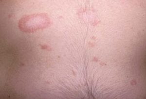
Distinguishing characteristics: Herald patch preceding annular plaques with collarette scale
Prurigo nodularis
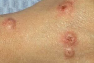
Distinguishing characteristics: Pruritic nodules, often on the extremities
Lichen planus can be diagnosed clinically in classic cases, although biopsy is often helpful to confirm the diagnosis and is required for more atypical presentations. A 4-mm punch biopsy should be adequate on the skin or in the mouth. The histology shows a characteristic “saw-tooth” pattern of epidermal hyperplasia; hyperparakeratosis with thickening of the granular cell layer; and vacuolar alteration of the basal layer of the epidermis, with an intense infiltration (mainly T cells) at the dermal-epidermal junction.
Reference:
- Usatine RP, Tinitigan M. Diagnosis and treatment of lichen planus. Am Fam Physician. 2011 Jul 1;84(1):53-60. PMID: 21766756.
Expert opinion – Dr. Ashish Shetty
Lichen planus is a chronic autoimmune inflammatory skin disorder characterized by pruritic, purple-colored, flat-topped papules commonly distributed over the wrists, forearms, and legs. The condition often heals with post-inflammatory pigmentation, which may take a long time to resolve. Common differential diagnoses include prurigo nodularis, papular urticaria, lichen simplex chronicus, psoriasis, and pityriasis rosea. Clinical examination usually allows differentiation from these conditions, and biopsy is only rarely required for confirmation. Topical tacrolimus and topical corticosteroids are effective in reducing both lesions and pruritus. In recalcitrant cases, short courses of oral tofacitinib or oral cyclosporine for 3 to 6 months may help in controlling symptoms.