

Pemphigus results from circulating immunoglobulin G (IgG) antibodies directed against desmosomes; these antibodies interfere with keratinocyte adhesion (Figure 1). Acantholysis occurs, resulting in the formation of bullae. There are 6 main types of pemphigus and their classification is based on the anatomic features of the lesion and the target antigens recognized by the autoantibodies. Eighty percent of patients with pemphigus have pemphigus vulgaris (PV). The annual incidence is estimated as one case per million population, but the condition is more common among Ashkenazi Jews and people of Mediterranean descent because of an association with certain human leukocyte antigen haplotypes.
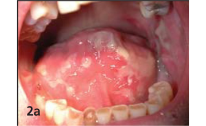
Differential diagnosis of pemphigus vulgaris
Mucous membrane pemphigoid
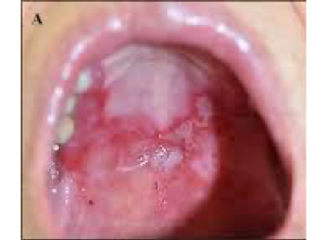
Distinguishing characteristics: Positive “clip sign” with linear deposits at the dermoepidermal junction demonstrated by direct immunofluorescence.
Erosive lichen planus
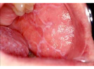
Distinguishing characteristics: Presence of Whickham’s striae adjacent to erosive lesions with a negative “clip sign.”
Bullous pemphigoid
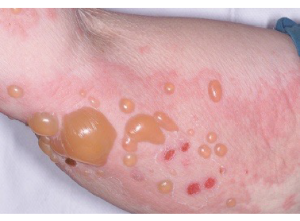
Distinguishing characteristics: Tense bullae or vesicles containing clear fluid, intense pruritus, symmetric distribution, and predilection for flexural areas and limb roots; mucosal involvement is uncommon.
Erythema multiforme
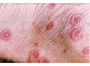
Distinguishing characteristics: Cutaneous rosette-like lesions composed of concentric zones with bullous centers and persistent pseudomembranes or crusting on the lips.
Pemphigus vulgaris can closely resemble several chronic erosive and bullous disorders, particularly those involving the oral mucosa. Careful evaluation of lesion morphology, associated systemic findings, histopathology, and immunofluorescence patterns is essential for accurate differentiation and timely initiation of therapy.
Reference:
- Ben Lagha N, Poulesquen V, Roujeau JC, et al. Pemphigus vulgaris: A case-based update. J Can Dent Assoc. 2005;71(9):667–672.
Expert opinion – Dr. Manjunath Hulmani
Pemphigus vulgaris is a recalcitrant and potentially debilitating disorder that must be carefully differentiated from other conditions that can clinically mimic oral pemphigus. Common differential diagnoses include oral lichen planus, mucous membrane pemphigoid, erythema multiforme, and Behçet’s disease. Clinical differentiation among these disorders can often be challenging; therefore, direct immunofluorescence (DIF) plays a crucial role in establishing the diagnosis. In pemphigus vulgaris, DIF typically demonstrates intercellular deposition of immunoreactants within the epidermis, producing the characteristic “fish-net” appearance. In contrast, mucous membrane pemphigoid shows a linear deposition of immunoreactants along the dermoepidermal junction (DEJ). Erosive lichen planus commonly demonstrates linear deposits of IgG, IgM, and C3 along the basement membrane zone (BMZ). Erythema multiforme is usually identified clinically by its characteristic targetoid cutaneous lesions, while DIF findings are generally negative. Similarly, Behçet’s disease does not show specific DIF positivity and is typically DIF-negative.