

Nummular dermatitis, also called discoid eczema or nummular eczema, is a pruritic eczematous dermatosis characterized by multiple coin-shaped lesions. This chronic inflammatory skin disease commonly involves the extremities and, less commonly, the trunk. Nummular dermatitis is regarded as a distinctive form of endogenous or idiopathic eczema, although some experts suggest it should be classified as a subtype of atopic dermatitis. Acutely, lesions may begin as papules or vesicles that coalesce into plaques. When established, lesions will appear symmetrically distributed, sharply defined, round or coin-shaped, and erythematous; eczematous plaques will range in size from 1 to 10 cm (Figure 1). Late-stage lesions may develop a drier scale and lichenification. Lesions may be associated with mild to intense pruritus. Lesion and symptom severity are exacerbated by behaviors that compromise the skin’s natural moisture barrier, such as harsh soaps and frequent, long, hot showers. The lower extremities are most commonly involved, followed by the upper extremities and trunk. The face and scalp are spared. Post inflammatory pigmentary changes typically persist after resolution. Dermoscopic findings may reveal scales, shiny yellow clots, and irregularly distributed brownish-red globules.
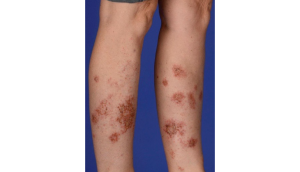
Differential diagnosis of nummular dermatitis
Asteatotic eczema
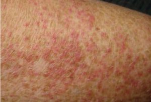
Distinguishing characteristics: Diffuse erythema and fine-scale, with small irregular fissures and cracks, most commonly on the bilateral lower legs
Fixed drug eruption
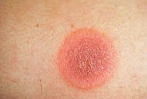
Distinguishing characteristics: Multiple well-circumscribed circular red to brown patches or edematous plaques that recur in the exact location when the patient is exposed to the implicated drug
Bullous pemphigoid
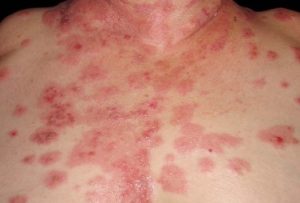
Distinguishing characteristics: Eczematous lesions for a prolonged period before the appearance of classic blisters
Majocchi granuloma
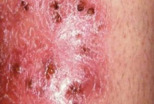
Distinguishing characteristics: Multiple perifollicular pustules coalescing to form erythematous scaly plaques
Recognizing the characteristic coin-shaped plaques of nummular dermatitis and distinguishing them from similar dermatoses is essential for accurate diagnosis and effective management.
Reference:
- Robinson CA, Love LW, Saleh HM, et al. Nummular Dermatitis. [Updated 2024 Mar 1]. In: StatPearls [Internet]. Treasure Island (FL): StatPearls Publishing; 2025 Jan-. Available from: https://www.ncbi.nlm.nih.gov/books/NBK565878/.
Expert opinion – Dr. Shashidhar T
The article on nummular dermatitis is scientifically accurate, clinically relevant, and closely aligned with real-world dermatology practice, particularly in the Indian OPD setting. Its description of coin-shaped, intensely pruritic plaques and the progression from papules and vesicles to scaling and lichenification reflects exactly what is commonly encountered in daily practice. Most patients I see are middle-aged or elderly, frequently with diabetes, xerosis, a history of hot-water bathing, or excessive soap use, and many present after inappropriate treatment with antifungal–steroid combination creams due to initial misdiagnosis as tinea. The differential diagnoses listed—asteatotic eczema, fixed drug eruption, bullous pemphigoid, and Majocchi granuloma—are precisely the conditions typically considered in clinical evaluation. Careful history-taking, morphological assessment, and selective investigations help in distinguishing these entities. The article rightly emphasizes barrier dysfunction and environmental triggers; however, in the Indian context, additional focus on soap misuse, topical steroid abuse, secondary infection from scratching, and high relapse rates due to inadequate long-term moisturization would further strengthen its practical value. With appropriate management—including strict soap avoidance, regular emollient use, correctly chosen topical steroids, and antihistamines—most patients experience 50–70% improvement within 7–10 days and near-complete resolution in 3–4 weeks, with residual pigmentation subsiding gradually over subsequent months. Overall, the article is dependable, practical, and well-suited for clinician education, patient counselling, and academic reference.