

Psoriasis is a chronic, autoimmune, relapsing skin disease of multifactorial etiology that affects about 2–3% of the general population. According to the authors, the prevalence of psoriasis is known in only one-fifth of all countries and is unevenly distributed in different geographic regions. Psoriasis occurs with equal frequency in both sexes, and its initial symptoms can manifest at any age; however, two peaks of incidence are noted. In men, this is at 30–39 years or 60–69 years, while in women, psoriasis can appear at 18–29 years or 50–59 years. The average age of its onset is 33 years. In the pediatric population, psoriasis is not common within this age group, with prevalence rates ranging from 0.13% in children aged 0–2 years to 0.67% in children aged 14–18 years. Clinically, several types of psoriasis are distinguished, but the most diagnosed is plaque psoriasis vulgaris (Figure 1).
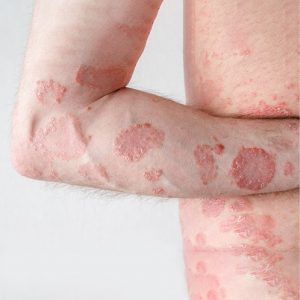
Differential diagnosis of psoriasis vulgaris
Differential diagnosis of psoriasis should be conducted in conjunction with all bacterial, viral diseases, tumors, precancerous lesions, mycosis fungoides, subacute lupus erythematosus, allergic and atopic dermatitis, lichen planus, and tinea pedis. In the differential diagnosis of psoriasis vulgaris, it is essential to consider five dermatological diseases.2
Nummular eczema
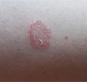
Distinguishing characteristics: Rounded, circular, desquamative erythematous lesions covered with vesicles, crusts, and scales, that are very itchy. Patients have either an atopic or an allergic diathesis. Epicutaneous allergy tests are frequently
Mycosis fungoides
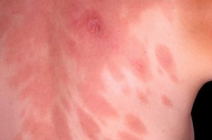
Distinguishing characteristics: Erythematous patches, little infiltrated and finely desquamating.2
Pityriasis rubra pilaris
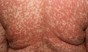
Distinguishing characteristics: Typical cases of follicular papules and infiltrating scales are observed, as well as typical hyperkeratosis.2
Duhring’s disease
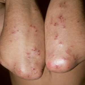
Distinguishing characteristics: Bilateral, symmetric localization on extensor surfaces of the limbs. Close-up observation will show papules and vesicles on the erythematous skin. In the eruptive phase, there are crusts filled with serum and blood, and lichenification due to scratching. In the chronic phase, this disease is constantly very itchy.2
Bowen’s disease
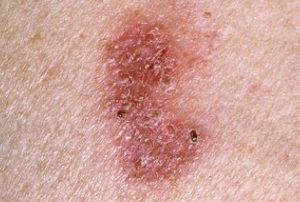
Distinguishing characteristics: Squamous cell carcinoma presenting inside the skin, erythematous, slightly infiltrated, finely desquamating, mainly single patches, showing no improvement with photo and local therapy.2
Differential diagnosis of psoriasis should be conducted in conjunction with all bacterial, viral diseases, tumors, precancerous lesions, mycosis fungoides, subacute lupus erythematosus, allergic and atopic dermatitis, lichen planus, and tinea pedis.2
Reference:
- Kowalska MK, Orłowska SM, Bednarczyk Ł, et al. Pathogenesis of psoriasis vulgaris and current management and therapeutic strategies including the role of emollients—A review of the current literature. Appl Sci. 2025;15:6811.
- Tuzun B. The differential diagnosis of psoriasis vulgaris. Pigmentary Disorders. 2016;3:3.
Expert opinion – Dr. Pawan Kumar
In the differential diagnosis of psoriasis vulgaris, several conditions may present with overlapping clinical features. Unlike the well-demarcated plaques of psoriasis, some mimickers display surfaces that are less uniform, with variable erythema and fissuring, particularly in flexural areas. Mycosis fungoides can resemble psoriasis but typically progresses through patch, plaque, and tumoral stages, with lesions showing fine scaling along with surface changes such as atrophy, telangiectasia, and mottled pigmentation; its distribution commonly involves the bathing-trunk region, including the buttocks, flanks, and inner thighs, with sparing of sun-exposed areas. Pityriasis rosea is characterized by an initial herald patch followed by multiple oval lesions aligned along Langer’s lines, creating a Christmas-tree pattern on the trunk, and its scale is thin, peripheral, and forms a collarette—features that help distinguish it from psoriasis. Subacute cutaneous lupus erythematosus (SCLE) may also mimic psoriasis, presenting with annular or polycyclic plaques on sun-exposed areas, marked photosensitivity, and a non-adherent trailing scale, healing with pigmentary changes. Seborrheic dermatitis, another common differential, presents with ill-defined erythematous patches covered by loose, greasy, yellowish scales, typically affecting seborrheic regions such as the scalp, nasolabial folds, and upper chest. Recognizing these characteristic differences is essential for accurate diagnosis and appropriate management.