

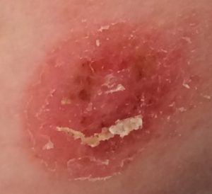
Pityriasis rosea is a self-limiting skin condition that presents as discrete scaly papules and plaques along the Langer lines (cleavage lines) over the trunk and limbs. The epidemiology and clinical course of pityriasis rosea suggest an infectious etiology. Temporal case clustering, which indicates infectious transmission, has been documented in regression analysis models. The diagnosis of pityriasis rosea is based on clinical and physical examination findings. Classic pityriasis rosea starts with a herald patch on the trunk (Figure 1) in up to 90% of cases. The patch is erythematous with slightly elevated scaling borders and a lighter depressed center. It can measure 3 cm or more in diameter and may be the only skin manifestation for approximately two weeks. The generalized rash, also known as the secondary eruption, presents on the trunk along the Langer lines and may extend to the upper arms and upper thighs. These lesions are smaller than the herald patch and can continue to appear for up to six weeks after the initial eruption.
Differential diagnosis of pityriasis rosea
Lichen planus
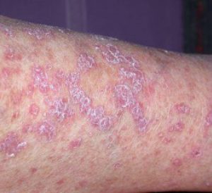
Distinguishing characteristics: 1- to 10-mm, sharply defined, flat-topped violaceous papules typically on wrists, lumbar region, shins, scalp, glans penis, and mouth; lesions may be asymptomatic
Nummular eczema
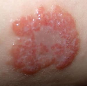
Distinguishing characteristics: Grouped small vesicles and papules 4 to 5 cm in diameter; round or coin-shaped lesions with an erythematous base and distinct borders, often on shins and the backs of hands; pruritus is often intense
Pityriasis lichenoides chronica
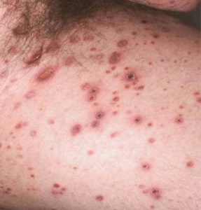
Distinguishing characteristics: Red-brown papules with central mica-like scales randomly arranged on trunk and proximal extremities with chronic, relapsing course; hypo- or hyperpigmentation may be present after lesions resolve
Pityriasis rosea–like eruption associated with medications
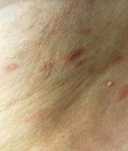
Distinguishing characteristics: Similar presentation to pityriasis rosea, but lesions resolve after causative medication is discontinued
The differential diagnosis of pityriasis rosea includes several conditions. If the diagnosis is uncertain, a skin biopsy will help exclude other pathologies. The histology of pityriasis rosea typically reveals focal parakeratosis, spongiosis, and acanthosis in the epidermis, along with extravasated red blood cells and perivascular infiltrates of lymphocytes, monocytes, and eosinophils in the dermis.
Reference:
- Villalon-Gomez JM. Pityriasis rosea: Diagnosis and treatment. Am Fam Physician. 2018;97(1):38–44.
Expert opinion – Dr. Ramesh Munshimappa
In its early or herald‐patch stage, pityriasis rosea can be mistaken for several conditions, including tinea corporis, annular psoriasis, annular lichen planus, lichen sclerosus et atrophicus, mycosis fungoides, and granuloma annulare. When pityriasis rosea progresses to its eruptive stage—where the lesions are smaller and distributed widely across the body—it may resemble other disorders such as drug exanthema, viral exanthema, secondary syphilis, guttate psoriasis, lichen planus, lichen nitidus, verruca plana, and pityriasis rubra pilaris. Recognizing these differential diagnoses at both stages is essential for accurate clinical assessment.