

Tinea capitis most commonly affects children. There are three types of tinea capitis: gray patch, black dot, and favus. Black dot, caused by Trichophyton tonsurans, is most common in the United States (Figure 1). Early disease can be limited to itching and scaling, but the more classic presentation involves one or more scaly patches of alopecia with hairs broken at the skin line (black dots) and crusting. Tinea capitis may progress to kerion, which is characterized by boggy, tender plaques and pustules. The child with tinea capitis will generally have cervical and suboccipital lymphadenopathy, and the physician may need to broaden the differential diagnosis if lymphadenopathy is absent. However, lymphadenopathy can also occur in nonfungal scalp disease, and the absence of lymphadenopathy in an otherwise typical presentation should not delay aggressive treatment for tinea capitis.
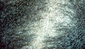
Differential diagnosis of tinea capitis
Psoriasis
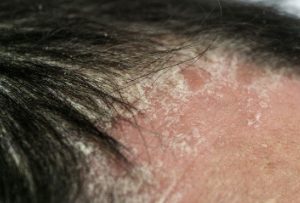
Distinguishing characteristics: Gray or silver scale; nail pitting; 70% of affected children have family history of psoriasis; involvement of other sites
Atopic dermatitis
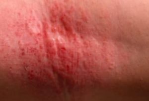
Distinguishing characteristics: Personal history or family history of atopy; less often annular; lymphadenopathy uncommon; alopecia less common
Bacterial scalp abscess
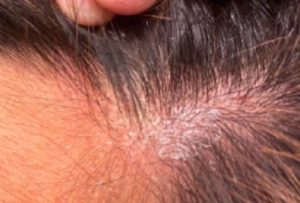
Distinguishing characteristics: Alopecia less likely; hair pluck is painful
Seborrheic dermatitis
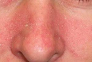
Distinguishing characteristics: Alopecia uncommon; lymphadenopathy uncommon; greasy scale; typical distribution involving nasolabial folds, hairline, eyebrows, postauricular folds, chest1
Trichotillomania
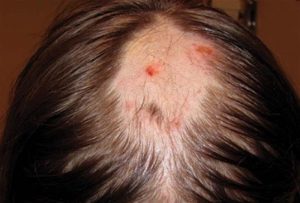
Distinguishing characteristics: No scale; commonly involves eyelashes and eyebrows; hairs of varying lengths
The most common mimic includes seborrheic dermatitis. In atypical cases, a KOH preparation can be performed by scraping the black dots (broken hairs) and looking for fungal spores. The spores of T. tonsurans will be contained within the hair shaft, but for the less common Microsporum canis, the spores will coat the outside of the hair shaft.
Reference:
- Ely JW, Rosenfeld S, Seabury Stone M. Diagnosis and management of tinea infections. Am Fam Physician. 2014;90(10):702–710.
Expert opinion – Dr. Syed Salahuddin
Alopecia areata typically presents as smooth, well-defined patches of hair loss with no scaling, no corkscrew hairs, and a negative KOH test. Syphilitic alopecia is characterized by a “moth-eaten” pattern of hair loss and is usually accompanied by other clinical signs of secondary syphilis. Trichotillomania shows hairs broken at varying lengths, absence of scaling, perifollicular hemorrhages, and characteristic findings such as hair dust and flame hairs. Seborrheic dermatitis and psoriasis usually present with diffuse scalp scaling but without significant inflammation.