

Tinea corporis (ringworm) typically presents as a red, annular, scaly, pruritic patch with central clearing and an active border (Figure 1). Lesions may be single or multiple, and their size generally ranges from 1 to 5 cm, but larger lesions and lesion confluence can also occur. Tinea corporis may be mistaken for many other skin disorders, especially eczema, psoriasis, and seborrheic dermatitis. A potassium hydroxide (KOH) preparation is often helpful when the diagnosis is uncertain based on history and visual inspection. Worsening after empiric treatment with a topical steroid should raise the suspicion of a dermatophyte infection. Conversely, if a nonfungal lesion is treated with an antifungal cream, it will likely not improve or may worsen. Cultures are usually unnecessary to diagnosing tinea corporis. A skin biopsy with periodic acid–Schiff (PAS) stain may be indicated in rare cases for atypical or persistent lesions.1
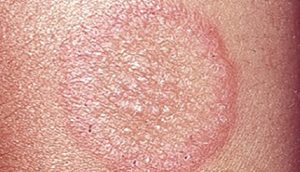
Differential diagnosis of tinea corporis
Annular psoriasis
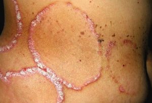
Distinguishing characteristics: Gray or silver scale; nail pitting; 70% of affected children have a family history of psoriasis1
Atopic dermatitis
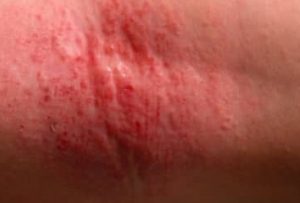
Distinguishing characteristics: Personal or family history of atopy; less likely to have active border with central clearing; lesions may be lichenified1
Granuloma annulare
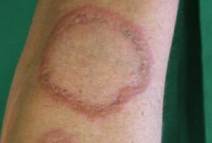
Distinguishing characteristics: No scale, vesicles, or pustules; nonpruritic; smooth; commonly on dorsum of hands or feet1
Seborrheic dermatitis
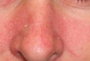
Distinguishing characteristics: Greasy scale on erythematous base with typical distribution involving nasolabial folds, hairline, eyebrows, postauricular folds, chest; annular lesions are less common1
Several diseases in the differential diagnosis can mimic tinea corporis, often presenting with annular lesions. Cases that do not respond to antifungal treatment or have a negative KOH microscopic examination should prompt further investigation. Clinicians must also consider more serious conditions, especially in cases with extensive skin involvement or severe disease.2
Reference:
-
- Ely JW, Rosenfeld S, Seabury Stone M. Diagnosis and management of tinea infections. Am Fam Physician. 2014;90(10):702–710.
Yee G, Syed HA, Al Aboud AM. Tinea Corporis. [Updated 2025 Feb 14]. In: StatPearls [Internet]. Treasure Island (FL): StatPearls Publishing; 2025 Jan-. Available from: https://www.ncbi.nlm.nih.gov/books/NBK544360/.
Expert opinion – Dr. Madura Chandu
Tinea corporis is a common fungal infection characterized by annular, erythematous, scaly plaques. While diagnosis is usually straightforward in classical presentations, atypical cases can be challenging and must be differentiated from conditions such as psoriasis, atopic eczema, granuloma annulare, and seborrhoeic dermatitis. Psoriasis typically shows thicker scales with a symmetrical distribution, whereas eczema presents as diffuse, pruritic lesions without an annular pattern. Granuloma annulare lacks scaling and often resolves spontaneously, and seborrhoeic dermatitis features greasy scales in sebaceous-rich areas. In contrast, tinea corporis demonstrates central clearing with active, scaly borders, and a KOH examination helps confirm the fungal etiology.