

Vitiligo is an acquired pigmentary skin disorder caused by the absence of pigmentary cells from the epidermis that results in white macules and patches on the body. The condition is usually associated with a few autoimmune disorders, with thyroid abnormalities being the most common ones. The etiology of vitiligo is unknown, but there are different theories to explain its pathogenesis. Vitiligo presents clinically with signs and symptoms of white spots on the body, distributed symmetrically, and more pronounced in individuals with dark skin. The lesions are characterized by well-demarcated, pearly white or depigmented macules and patches, which may be oval, round, or linear in shape, with convex borders that range in size from a few millimeters to centimeters and are enlarge centrifugally. There are different clinical variants of vitiligo, including trichrome, marginal inflammatory, and quadrichrome vitiligo. Initial lesions occur most frequently on the hands, forearms, feet, and face, favoring a periocular or perioral distribution (Figure 1).1
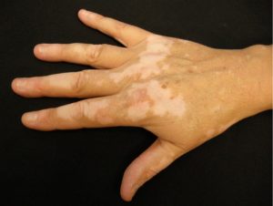
Differential diagnosis of vitiligo
Halo nevus
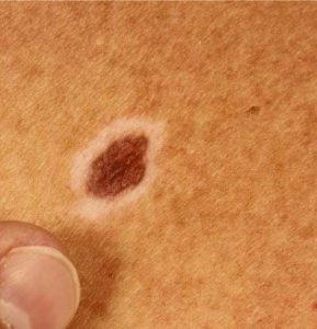
Distinguishing characteristics: It is a type of melanocytic nevus surrounded by an oval halo of depigmentation1
Progressive macular hypomelanosis
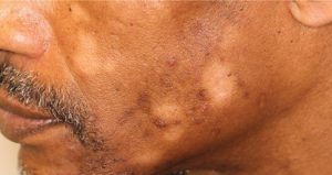
Distinguishing characteristics: It clinically presents as asymptomatic hypopigmented patches in young adults1
Drug-induced leukoderma
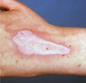
Distinguishing characteristics: Potent topical or intralesional corticosteroids can induce hypopigmentation at the site of application. Depigmentation is also noticed in patients treated with the tyrosine kinase inhibitors and epidermal growth factor receptor inhibitors.1
Hypopigmented mycosis fungoides
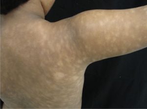
Distinguishing characteristics: It is a variant of early-stage MF commonly seen in children and dark-skinned people. It appears as widespread hypopigmented patches with atrophy and scaling and atrophy.1
Many other skin disorders present with hypopigmentation or depigmentation and must be distinguished to determine the right diagnosis, advise the patient on prognosis, and prescribe the correct treatment.2
Reference:
- Ahmed jan N, Masood S. Vitiligo. [Updated 2023 Aug 7]. In: StatPearls [Internet]. Treasure Island (FL): StatPearls Publishing; 2025 Jan-. Available from: https://www.ncbi.nlm.nih.gov/books/NBK559149/.
- Goh BK, Pandya AG. Presentations, signs of activity, and differential diagnosis of vitiligo. Dermatol Clin. 2017;35(2):135–144.
Expert opinion – Dr. Vinayak Venkatesh
I would like to add a few more conditions that may mimic vitiligo. These include depigmentation secondary to intra-articular or intralesional triamcinolone injections, depigmentation following superficial burns, achromic naevus, depigmentation on the scalp due to hair-dye reactions, and depigmentation resulting from bindi dermatitis. These are some of the conditions I have encountered in my clinical practice.